Skeda:Anaplastic astrocytoma.jpg
Kërceni tek navigimi
Kërceni tek kërkimi
Madhësia e këtij shikimi: 800 × 552 pixel. Rezolucione të tjera: 320 × 221 pixel | 640 × 442 pixel | 1.024 × 707 pixel | 1.200 × 828 pixel.
Skedari origjinal (1.200 × 828 pixela, madhësia e skedës: 200 KB, tipi MIME: image/jpeg)
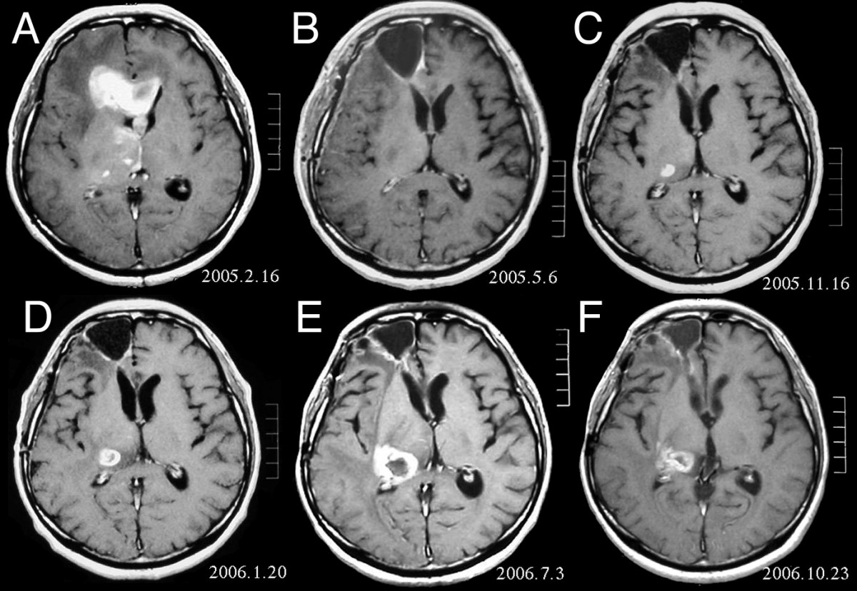
| Përshkrimi | MRI of brain. (A) Initial MRI on February 16, 2005, shows a tumor in the right and left frontal lobe as well as the right thalamus. (B) MRI after surgery, radiation and chemotherapy. The tumor has completely disappeared except for slight enhancement adjacent to the surgical margin. (C) Recurrence of the thalamic tumor despite maintenance chemotherapy on November 16, 2005. (D) Increase in size of the thalamic tumor two months after stereotactic radiotherapy. (E) After 6 cycles of TMZ therapy, the thalamic lesion enlarged, and the patient developed dysarthria and hemiparesis. (F) After 2 courses of treatment with interferon-beta and TMZ, the tumor shows a partial response. |
| Data | |
| Burimi | Fujimaki T, Ishii H, Matsuno A, Arai H, Nakagomi T.Effectiveness of interferon-beta and temozolomide combination therapy against temozolomide-refractory recurrent anaplastic astrocytoma.World J Surg Oncol. 2007 Aug 4;5:89. PMID 17683572 doi:10.1186/1477-7819-5-89 |
| Autori | Fujimaki T, Ishii H, Matsuno A, Arai H, Nakagomi T. |
| Leja (Duke ripërdorur këtë skedar) |
BioMedCentral License |
Kjo skedë është dhënë për përdorim sipas licensës Creative Commons Attribution 2.0 Generic.
- Je i lirë të:
- ta shpërndani – ta kopjoni, rishpërndani dhe përcillni punën
- t’i bëni “remix” – të përshtatni punën
- Sipas kushteve të mëposhtme:
- atribuim – Duhet t’i jepni meritat e duhura, të siguroni një lidhje për tek licenca dhe të tregoni nëse janë bërë ndryshime. Këtë mund ta bëni në ndonjë mënyrë të arsyeshme, por jo në ndonjë mënyrë që sugjeron se licencuesi ju del zot juve apo përdorimit tuaj.
Historiku i skedarit
Kliko mbi një datë/orë për ta parë skedarin siç është shfaqur në atë kohë.
| Data/Ora | Miniatura | Përmasat | Përdoruesi | Koment | |
|---|---|---|---|---|---|
| e tanishme | 25 shkurt 2008 17:47 | | 1.200 × 828 (200 KB) | wikimediacommons>Filip em | {{Information |Description=MRI of brain. (A) Initial MRI on February 16, 2005, shows a tumor in the right and left frontal lobe as well as the right thalamus. (B) MRI after surgery, radiation and chemotherapy. The tumor has completely disappeared except f |
Përdorimi i skedarit
faqe lidhet tek kjo skedë: